Echocardiography is a test that uses sound waves to produce live images of your heart.The image is an echocardiogram. This test allows your doctor to monitor how your heart and its valves are functioning.
The images can help them spot: blood clots in the heart fluid in the sac around the heart problems with the aorta, which is the main artery connected to the heart An echocardiogram is key in determining the health of the heart muscle,
Types of Echocardiogram
- Transthoracic echocardiography
- Transesophageal echocardiography
- Stress echocardiogram
- Three-dimensional echocardiography
- Fetal echocardiography
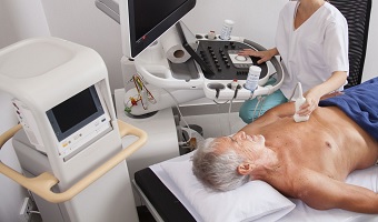
Echocardiograms are performed by placing a transducer on the chest and aiming it at the heart. The transducer transmits and receives sound waves that bounce off the heart. A computer compiles these returning sound waves, or echoes, and turns them into a picture of the heart.
What can detect after Echocardiogram
- damage to the heart muscle
- heart defects
- heart size
- pumping strength
- valve problems